
DMSA Case 1
Details on the Request Card
24yr old female with recurrent UTIs. ? renal scarringProcedure
Anterior and posterior images are shown. Split function is R - 53%, L - 47%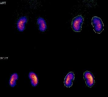
Questions
1) Name the radiopharmaceutical and isotope
2) How long from injection to imaging?
3) Why?
4) What is the accepted range for normal split function?
5) What are the green and white lines on the right hand images?
6) What percentage of the radiopharmaceutical is excreted?
The text is entirely the opinion of the author and does not necessarily reflect that of RUH NHS Trust or the Bristol Radiology Training Scheme. Website content devised by Paul McCoubrie.